immunofixation electrophoresis principle
Immunofixation electrophoresis is the study of protein antigens and their split products and evaluation of myeloma. Grabar and Williams first coined the term immunoelectrophoresis in 1953.
Ppt Electrophoresis And Its Application For Diagnosis Powerpoint Presentation Id 3498074
The distances that individual proteins travel depend on their size shape and electrical charge.

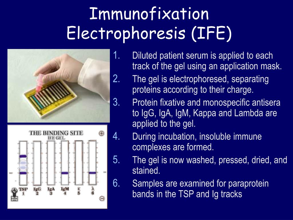
. 1 Serum or urine immunofixation negative for a monoclonal protein or a polyclonal pattern is considered to be normal. This was described in 1964. After application of an electric current that allows the separation of proteins according to their size antibodies specific for each type of immunoglobulin are laid upon the gel.
A polyclonal immunoglobulin pattern in the serum or urine. After the separation of serum and urine proteins according to their charge the gel is incubated with different specific antisera. It is a combination of two processes electrophoresis and immuno-diffusion.
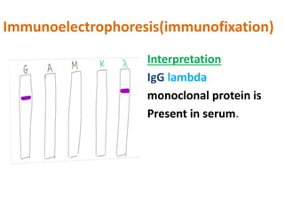
The reaction can be detected by visual inspection in indirect light by protein. Typically this testing determines the presence and type of monoclonal proteins eg IgG kappa. In some pathological cases multiple myeloma etc abnormal monoclonal bands appear in electrophorogram.
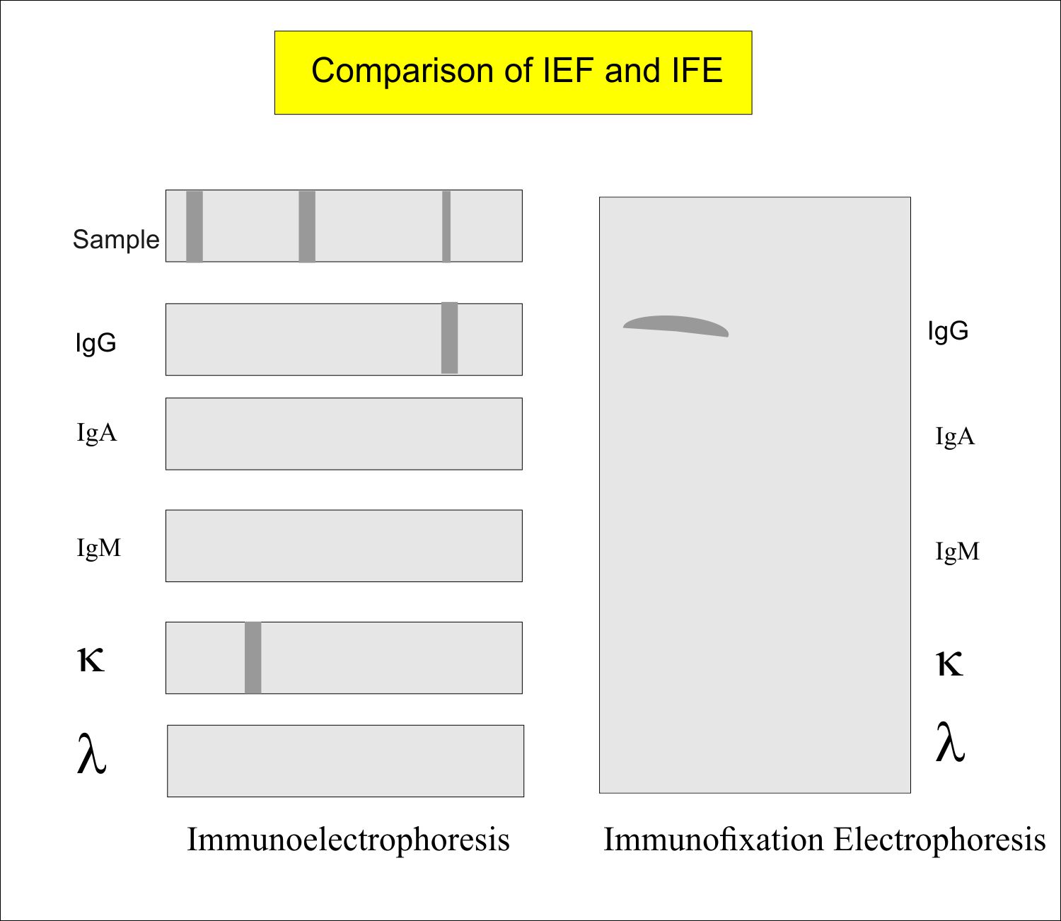
Certain diseases result in the growth of an excess number of antibody-producing cells. An antigen mixture is first separated into its component parts by electrophoresis and then tested by double immuno-diffusion. Immunofixation offers the worker an economical means of physically locating a protein in an electrophoretic strip and is ideally suited to forensic medicine genetic studies or research.
An immunofixation blood test also known as protein electrophoresis measures certain proteins in the blood. Immunofixation electrophoresis consists of an electrophoretic phase followed by a fixation phase in which antiserum is used to precipitate the protein. Immunofixation consists of an electrophoresis phase and a fixation phase.
The principle is the same in both processes. The band then glows. This is accomplished by a method known as immunofixation electrophoresis IFE IFE consists of applying a fluorescent antibody against a constituent directly to an electrophoresis gel.
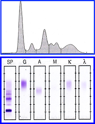
The method is as simple and economical as the commonly used one- or two-dimensional immunoelectrophoresis yet yields considerably more information. Immunofixation electrophoresis 172 is the most sensitive method for identifying and characterizing M proteins. Immunofixation electrophoresis IFE is a technique for the identification of proteins within complex mixtures after separation by either conventional zone electrophoresis or isoelectric focusing.
Hellabio Immunofixation Electrophoresis IFE kits are intended for in vitro diagnosis of monoclonal paraproteins in human serum and other biological samples. As long as the antibody is in slight excess or near equivalency the antigenantibody complex remains insoluble. It needs antibodies also known as immunoglobulins to react with proteins for characterization and separation.
A trough is then cut in the gel into which antibodies are placed. Electrophoresis separates the proteins in the specimen according to net charge. The lab treats the gel to keep only certain proteins.
In some diseases these cells can produce a large number of antibodies that are all exactly the same. Immunofixation as immunoelectrophoresis takes place in two steps. The identification of these monoclonal bands can be done by different immunological techniques.
It is a process of a combination of immuno-diffusion and electrophoresis. The technique consists of depositing a serum or urine which has been previously concentrated sample on a gel. This test uses an electric current to push proteins in a urine sample through a special gel.
Proteins play many important roles including providing energy for the body rebuilding muscles and supporting the immune system. Immunofixation should be used to confirm the presence of an M protein and to identify the heavy-chain type and the light-chain class. During the test an electric current is used to move the proteins across a thin layer of agarose gel.
Immunofixation is an immunological method for detection and identification of monoclonal proteins in serum and urine. 1 In the former the serum is applied to an agarose. First six aliquots of the test specimen are applied to an agarose gel.
CSF is the fluid that surrounds your brain and spinal cord. 1 Serum protein electrophoresis should be performed whenever MM WM or AL is suspected and if clinical suspicion is high immunofixation studies should be done despite a normal serum electrophoretic pattern. Immunofixation involves two steps.
Most commonly antigens which are often immunoglobulins are separated by electrophoresis followed by precipitation with specific antibodies in situ. Answer Immunofixation consists of an electrophoresis phase and a fixation phase. There are two main types of proteins in the blood.
Normally your CSF does not contain much protein. Immunoelectrophoresis is simply agars precipiration in an electric field. These studies are also.
Immunofixation electrophoresis is replacing the electrophoresis because of its rapidity and ease of interpretation. The gel is then processed in order to remove the antisera excess prior to the final staining step. It thus appears to be more or less narrow bands on the gel which are at different immunoglobulins.
An increase in the amount of these proteins could be a sign of an inflammatory or immune disorder. If your body makes M-protein it may mean that you have one of several types of cancer such as multiple myeloma or another serious health problem. Protein electrophoresis is a method for separating the proteins found in blood serum or urine.
Immunofixation electrophoresis or immunosubtraction capillary electrophoresis identifies the type of immunoglobulin protein s present as monoclonal bands on a protein electrophoresis pattern. This test uses an electrical current on a CSF sample to separate out types of protein called immunoglobulins. The specimen to be tested can be serum urine or other body fluids.
Antigens are placed into wells cut in a gel without antibody and electrophoresed. CSF immunofixation that does not reveal oligoclonal bands is also considered normal. If the constituent is present the antibody will stick to it and survive without being washed away.

High Resolution Electrophoresis And Immunofixation Techniques And Interpretation Kindle Edition By Keren David F Professional Technical Kindle Ebooks Amazon Com

Serum Protein Electrophoresis Ppt Video Online Download

Hemoglobin Electrophoresis Capillary Electrophoresis Physical Properties Electrode Gel

Electropherograms On Capillary Electrophoresis Of Paraprotein Igm A Download Scientific Diagram
Sd Electrophoresis And Immunofixation
Protein Electrophoresis And Mass Spectrometry Of Serum And Download Scientific Diagram

Electrophoresis Principle Ppt Video Online Download
View Case

Electropherograms On Capillary Electrophoresis Of Paraprotein Igm A Download Scientific Diagram

Gel Electrophoresis Types Principles Instrumentation And Applications Gel Protein Biology Principles
Sd Electrophoresis And Immunofixation

Electrophoresis Immunoelectrophoresis And Immunofixation Youtube

Electrophoresis Immunoelectrophoresis And Immunofixation Youtube

Serum Protein Electrophoresis Definition Principle Types Procedure Results Youtube

Serum Protein Electrophoresis And Multiple Myeloma With Link To And Excerpts From Screening Panels For Detection Of Monoclonal Gammopathies Tom Wade Md

Interpreting Serum Protein Electrophoresis
Monoclonal Immunoglobulin Ig Monoclonal Antibody Immunofixation Electrophoresis Ife Labpedia Net

Agarose Gel Electrophoresis Youtube

Immunofixation An Overview Sciencedirect Topics


Comments
Post a Comment